Followers

Wednesday 12 May 2021

ARTERIAL BLOOD PRESSURE

 DEFINITIONS AND NORMAL VALUES

  Arterial blood pressure is defined as the lateral pres sure exerted by the column of blood on wall of arteries. The pressure is exerted when blood flows through the arteries Generally, the term blood pressure refers to arterial blood pressure.

Arterial blood pressure is expressed in four terms

1.Systolic blood pressure                        2.Diastolic blood pressure.                                    3. Pulse pressure.                                                      4.Mean arterial blood pressure.


SYSTOLIC BLOOD PRESSURE                                    Systolic blood pressure (systolic pressure) is defined as the maximum pressure exerted in the arteries during systole of heart

Normal systolic pressure:

 120 mm Hg (110 to 140 mm Hg)


DIASTOLIC BLOOD PRESSURE

Diastolic blood pressure (diastolic pressure) is defined as the minimum pressure exerted in the arteries during diastole of heart


Normal diastolic pressure:

80 mm Hg (60 to 80 mm Hg)


PULSE PRESSURE

Pulse pressure is the difference between the systolic pressure and diastolic pressure


Normal pulse pressure:

40 mm Hg (120 - 80 = 40)


MEAN ARTERIAL BLOOD PRESSURE

Mean arterial blood pressure is the average pressure existing in the arteries. It is not the arithmetic mean of systolic and diastolic pressures. It is the diastolic pressure plus one third of pulse pressure. To determine the mean pressure, diastolic pressure is considered than the systolic pressure. It is because the diastolic period of cardiac cycle is longer (0.53 second) than the systolic period (0.27 second)


Normal mean arterial pressure:

93 mm Hg (80+ 13 = 93).


Formula to calculate mean arterial blood pressure Mean arterial blood pressure

 =Diastolic pressure 1/3 of pulse pressure

 = 80 +40 3 = 93.3 mm Hg


VARIATIONS

Blood pressure is altered in physiological and pathological conditions Systolic pressure is subjected for variations easily and quickly and its variation occurs in a wider range. Diastolic pressure is not subjected for easy and quick variations and its variation occurs in a narrow range.


PHYSIOLOGICAL VARIATIONS

1. Age

Arterial blood pressure increases as age advances. Table 100.1 shows systolic and diastolic pressures at different age.


2. Sex

In females, up to the period of menopause, arterial pressure is 5 mm Hg, less than in males of same age. After menopause, the pressure in females becomes equal to that in males of same age.


3. Body Built

Pressure is more in obese persons than in lean persons.


4. Diurnal Variation

In early morning, the pressure is slightly low. It gradually increases and reaches the maximum at noon. It becomes low in evening.

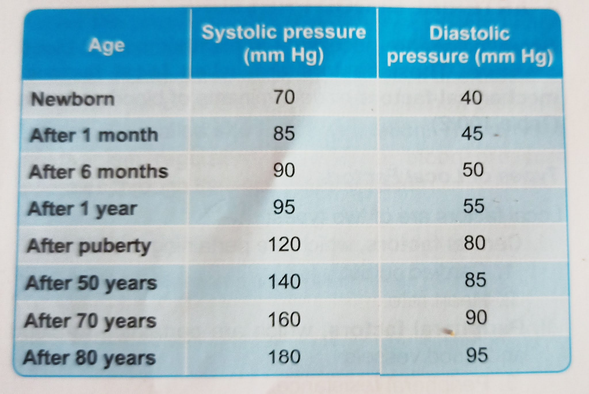
Arterial blood pressure in different age

5. After Meals

Arterial blood pressure is increased for few hours after meals due to increase in cardiac output.


6. During Sleep

Usually, the pressure is reduced up to 15 to 20 mm Hg during deep sleep. However, it increases slightly during sleep associated with dreams


7. Emotional Conditions

During excitement or anxiety, the blood pressure is increased due to release of adrenaline


8. After Exercise

After moderate exercise, systolic pressure increases by 20 to 30 mm Hg above the basal level due to increase in rate and force of contraction and stroke volume Normally, diastolic pressure is not affected by moderate exercise. It is because, the diastolic pressure depends upon peripheral resistance, which is not altered by moderate exercise.

  After severe muscular exercise, systolic pressure rises by 40 to 50 mm Hg above the basal level. But the diastolic pressure reduces because the peripheral resistance decreases in severe muscular exercise. More details are given in Chapter 114


PATHOLOGICAL VARIATIONS

Pathological variations of arterial blood pressure are hypertension and hypotension. Refer applied physiology of this chapter for details.


DETERMINANTS OF ARTERIAL BLOOD PRESSURE: FACTORS MAINTAINING ARTERIAL BLOOD PRESSURE:

Some factors are necessary to maintain normal blood pressure. These factors are called local factors, mechanical factors or determinants of blood pressure . 


Types of Local Factors

Local factors are of two types:                                1. Central factors, which are pertaining to the heart

1. Cardiac output.                                                    2. Heart rate.

II. Peripheral factors, which are pertaining to blood and blood vessels:

3. Peripheral resistance.                                          4. Blood volume                                                        5. Venous return.

6. Elasticity of blood vessels.

7. Velocity of blood flow.                                          8. Diameter of blood vessels.

9. Viscosity of blood.


CENTRAL FACTORS

1. Cardiac Output

   Cardiac output is the important factor which maintains systolic blood pressure. Systolic pressure is directly proportional to cardiac output. Whenever the cardiac output increases, the systolic pressure is increased and when cardiac output is less, the systolic pressure is reduc Cardiac output increases in muscular exercise, emotional conditions, etc. So, in these conditions, the systolic pressure is increased. In conditions like myocardial infarction, the cardiac output decreases, resulting in fall in systolic pressure.


2. Heart Rate

Moderate changes in heart rate do not affect arterial blood pressure much. However, marked alteration in the heart rate affects the blood pressure by altering cardiac output. 


PERIPHERAL FACTORS

3. Peripheral Resistance

 Peripheral resistance is the important factor, which maintains diastolic pressure. Diastolic pressure is directly proportional to peripheral resistance Peripheral resistance is the resistance offered to the blood flow at the periphery. Resistance is offered at arterioles, which are called the resistant vessels. When peripheral resistance increases, diastolic pressure is increased and when peripheral resistance decreases, the diastolic pressure is decreased.


Local factor determining arterial blood pressure. 

4. Blood Volume

Blood pressure is directly proportional to blood volume. Blood volume maintains the blood pressure through the venous return and cardiac output. If the blood volume increases, there is an increase in venous return and cardiac output, resulting in elevation of blood pressure. 


5. Venous Return

Blood pressure is directly proportional to venous return. When venous return increases, there is an in crease in ventricular filling and cardiac output, resulting in elevation of arterial blood pressure.


6. Elasticity of Blood Vessels

Blood pressure is inversely proportional to the elasticity of blood vessels. Due to elastic property, the blood vessels are distensible and are able to maintain the pressure. When the elastic property is lost, the blood vessels become rigid (arteriosclerosis) and pressure increases as in old age. Deposition of cholesterol, fatty acids and calcium ions produce rigidity of blood vessels and atherosclerosis, leading to increased blood pressure.


7. Velocity of Blood Flow

Pressure in a blood vessel is directly proportional to the velocity of blood flow. If the velocity of blood flow increases, the resistance is increased. 

Effect of blood volume and venous return on arterial blood pressure. 

8. Diameter of Blood Vessels

Arterial blood pressure is inversely proportional to the diameter of blood vessel. If the diameter decreases, the peripheral resistance increases, leading to increase in the pressure.

9. Viscosity of Blood

Arterial blood pressure is directly proportional to the viscosity of blood. When viscosity of blood increases, the frictional resistance is increased and this increases the pressure.


REGULATION OF ARTERIAL BLOOD PRESSURE

Arterial blood pressure varies even under physiological conditions. However, immediately it is brought back to normal level because of the presence of well-organized regulatory mechanisms in the body. Body has four such regulatory mechanisms to maintain the blood pressure within normal limits:

I. Nervous mechanism or short-term regulatory mech anism. II. Renal mechanism or long-term regulatory mechanism.

III. Hormonal mechanism.

IV. Local mechanism.

BLOOD GROUP

 INTRODUCTION

  When blood from two individuals, sometimes agglutination (clumping) of RBCs occurs. Agglutination is because of the immunological reactions. But why it occurs in some cases and not in other cases remained a mystery until the discovery of blood groups by the Austrian Scientist Karl Landsteiner, in 1901. He was honored with Nobel Prize in 1930 for this discovery.

ABO BLOOD GROUPS

Determination of ABO blood groups depends upon the immunological reaction between antigen and antibody. Landsteiner found two antigens on the surface of RBCs and named them as A antigen and B antigen. These antigens are also called agglutinogens because of their capacity to cause agglutination of RBCs. He noticed the corresponding antibodies or agglutinins in the plasma and named them anti-A or a-antibody and anti-3 or B-antibody. However, a particular agglutinogen and the corresponding agglutinin cannot be present together. If present, it causes clumping of the blood. Based on this, Karl Landsteiner classified the blood groups. Later it became the 'Landsteiner's Law' for grouping the blood.


LANDSTEINER'S LAW

Landsteiner's law states that:

  1. If a particular agglutinogen (antigen) is present in the RBCs of a person, corresponding agglutinin (antibody) must be absent in the serum.

2. If a particular agglutinogen is absent in the FBC the corresponding agglutinin must be present in s serum.

Though the second part of Landsteiner's law is a fact, it is not applicable to Rh factor.

BLOOD GROUP SYSTEMS

More than 20 genetically determined blood group systems are known today. But Landsteiner discovered two blood group systems called the ABO system and the Rh system. These two blood group systems are the most important ones that are determined before blood transfusions.

ABO SYSTEM

Based on the presence or absence of antigen A and antigen B, blood is divided into four groups:

1. 'A' group.

2. B' group.

3. 'AB' group.

4. 'O' group.

 Blood having antigen A belongs to 'A' group. This blood has B-antibody in the serum. Blood with antigen B and a-antibody belongs to 'B' group. If both the antigens are present, blood group is called 'AB' group and serum of this group does not contain any antibody. If both anti gens are absent, the blood group is called 'O' group and both and B antibodies are present in the serum.

 Antigens and antibodies present in different groups of ABO system are given in Table a Percentage of people among Asian and European population belonging to different blood group is given in Table b. 


Table a. Antigen and antibody present in ABO blood groups. 

Table b. Percentage of people having different blood groups. 


DETERMINATION OF ABO GROUP
Determination of the ABO group is also called bl grouping, blood typing or blood matching

Principle of Blood Typing: Agglutination
 Blood typing is done on the basis of apgulinssion: Agglutination means the collection of separate particles like RBCs into clumps or masses Agglutination occurs if an antigen is mixed with its corresponding and body which is called isoagglutinin. Agglutination occurs. when A antigen is mixed with anti-A or when B antigen is mixed with anti-B.

Requisites for Blood Typing
 To determine the blood group of a person, a suspension of his/her RBC and testing antisera are required. Suspension of RBC is prepared by mixing blood drops with isotonic saline (0.9%)

Test sera are:
 1. Antiserum A, containing anti-A or a-antibody 2. Antiserum B. containing anti-B or B-antibody

Procedure
 1. One drop of antiserum A is placed on one end of a glass slide (or a tile) and one drop of antiserum B on the other end.
 2. One drop of RBC suspension is mixed with each antiserum. The slide is slightly rocked for 2 minutes.The presence or absence of agglutination is observed by naked eyes and if necessary, it is confirmed by using microscope.
 3. Presence of agglutination is confirmed by the presence of thick masses (clumping) of RBCs, 4. Absence of agglutination is confirmed by clear mixture with dispersed RBCs.       

Results
 1. If agglutination occurs with antiserum A
 The antiserum A contains a-antibody. The agglutination occurs if the RBC contains A antigen.So, the blood group is A. 
 2.If agglutination occurs with antiserum B The antiserum B contains B-antibody. The agglutination occurs if the RBC contains B antigen So, the blood group is B
 3. If agglutination occurs with both antisera A and B The RBC contains both A and B antigens to cause agglutination And, the blood group is AB.
Determination of blood group. 
4.if agglutination does not occur either with antiserum A Or antiserum B:
The agglutination does not occur because RBC does not contain any antigen. 
                                                                     IMPORTANCE OF ABO GROUPS IN BIOOD TRANSFUSION

During blood transfusion, only compa lood must be used. The one who gives blood is called 'donor' and the one who receives the blood is called 'recipient'.
  While transfusing the blood, antigen of the donor and the antibody of the recipient are considered. The antibody of the donor and antigen of the recipient are ignored mostly.
  Thus, RBC of 'O' group has no antigen and so agglutination does not occur with any other group of blood. So, 'O' group blood can be given to any blood group persons and the people with this blood group are called 'universal donors'.
  Plasma of AB group blood has no antibody. This does not cause agglutination of RBC from any other group of blood. People with AB group can receive blood from any blood group persons. So, people with this blood group are called 'universal recipients'.

MATCHING AND CROSSMATCHING

Blood matching (typing) is a laboratory test done to determine the blood group of a person. When the person needs blood transfusion, another test called crossmatching is done after the blootd is typed. It is done to find out whether the person's body will accept the donor's blood or not.

For blood matching, RBC of the individual (recipient) and test sera are used. Crossmatching is done by mising the serum of the recipient and the RBCs of don Crossmatching is always done before blood transfun If agglutination of RBCs from a donor occurs d crossmatching, the blood from that person is not for transfusion. 9 d

Matching= Recipient's RBC Crossmatching = Recipient's serum + Donor's RC+ Test sera

INHERITANCE OF ABO AGGLUTINOGENS AND AGGLUTININS
  Blood group of a person depends upon the two ge es inherited from each parent. Gene A and gene Bre dominant by themselves and gene O is recessive. Inheritance of blood group is represented schematically as given in Table c. 
   Agglutinogens appear during the 6th month of fetal life. Concentration at birth is 1/5 of the adult concentra tion. It rises to the adult level at puberty. Agglutinogens are present not only in RBCs but also present in many organs like salivary glands, pancreas, kidney, liver, lungs, etc. The A and B agglutinogens are inherited from the parents as Mendelian phenotypes.
  Agglutinin a or B is not produced during fetal life. It starts appearing only 2 or 3 months after birth. Agglutinin is produced in response to A or B agglutinogens which enter the body through respiratory system or digestive system along with bacteria. Agglutinins are the gamma globulins which are mainly IgG and IgM immunoglobu lins.
Inheritance of ABO blood group. 

Rh FACTOR
  Rh factor is an antigen present in RBC. This antigen was discovered by Landsteiner and Wiener. It was first discovered in Rhesus monkey and hence the name 'Rh factor'. There are many Rh antigens but only the D antigen is more antigenic in human.
  The persons having D antigen are called 'Rh positive' and those without D antigen are called 'Rh negative". Among Indian pop ation, 85% of people are Rh positive and 15% are Rh negative. Percentage of Rh positive people is more among black people.
  Rh group system is different from ABO group system because, the antigen D does not have corresponding natural antibody (anti-D). However, if Rh positive blood is transfused to a Rh negative person anti-D is developed in that person. On the other hand, there is no risk of complications if the Rh positive person receives Rh negative blood.

INHERITANCE OF Rh ANTIGEN
 Rhesus factor is an inherited dominant factor. It may be homozygous Rhesus positive with DD or heterozygous Rhesus positive with Dd  Rhesus negative occurs only with complete absence of D (i.e. with homozygous dd).

APPLIED PHYSIOLOGY

  TRANSFUSION REACTIONS DUE TO ABO INCOMPATIBILITY
  Transfusion reactions are the adverse reactions in the body, which occur due to transfusion error that involves transfusion of incompatible (mismatched) blood. The reactions may be mild causing only fever and hives (skin disorder characterized by itching) or may be severe lead ing to renal failure, shock and death.
   In mismatched transfusion, the transfusion reactions occur between donor's RBC and recipient's plasma. So, if the donor's plasma contains agglutinins against recipient's RBC, agglutination does not occur because these antibodies are diluted in the recipient's blood.
   But, if recipient's plasma contains agglutinins against donor's RBCs, the immune system launches a response against the new blood cells. Donor RBCs are agglutinated resulting in transfusion reactions.

ERYTHROPOIESIS

 DEFINITION:

   Erythropoiesis is the process of the origin, development and maturation of erythrocytes. Hemopoiesis or hematopoiesis is the process of origin, development and maturation of all the blood cells.


SITE OF ERYTHROPOIESIS:

IN FETAL LIFE

 in fetal life, the erythropoiesis occurs in three stages. 

1. Mesoblastic Stage

During the first 2 months of intrauterine life, the RBCs are produced from mesenchyme of yolk sac.

2. Hepatic Stage

From third month of intrauterine life, liver is the main organ that produces RBCs. Spleen and lymphoid organs are also involved in erythropoiesis.

3. Myeloid Stage

During the last 3 months of intrauterine life, the RBCs are produced from red bone marrow and liver.

IN NEWBORN BABIES, CHILDREN AND ADULTS

  in newborn babies, growing children and adults, RBCs are produced only from the ted bone marrow. 

1. Up to the Age of 20 Years                                 RBCs are produced from red bone marrow of all bones (long bones and all the flat bones).

2. After the Age of 20 Years

RBCs are produced from membranous bones like vertebra, sternum, ribs, scapula, iliac bones and skull bones and from the ends of long bones. After 20 years of age, the shaft of the long bones becomes yellow bone marrow because of fat deposition and loses the erythropoietic function.

In adults, liver and spleen may produce the blood cells if the bone marrow is destroyed or fibrosed. Collectively bone marrow is almost equal to liver in size and weight. It is also as active as liver. Though bone marrow is the site of production of all blood cells, comparatively 75% of the bone marrow is involved in the production of leukocytes and only 25% is involved in the production of erythrocytes.

  But still, the leukocytes are less in number than the erythrocytes, the ratio being 1:500. This is mainly because of the lifespan of these cells. Lifespan of erythrocytes is 120 days whereas the lifespan of leukocytes is very short ranging from 1 to 10 days. So, the leukocytes need larger production than erythrocytes to maintain the required number.


PROCESS OF ERYTHROPOIESIS

STEM CELLS

Stem cells are the primary cells capable of self-renewal and differentiating into specialized cells (Chapter 1). Hematopoietic stem cells are the primitive cells in the bone marrow, which give rise to the blood cells.

Hematopoietic stem cells in the bone marrow are called uncommitted pluripotent hematopoietic stem cells (PHSC). PHSC is defined as a cell that can give rise to all types of blood cells. In early stages, the PHSC are not designed to form a particular type of blood cell. And it is also not possible to determine the blood cell to be de veloped from these cells, hence, the name uncommitted PHSC . In adults, only a few of these cells are present. But the best source of these cells is the umbilical cord blood.

  When the cells are designed to form a particular type of blood cell, the uncommitted PHSCS are called committed PHSCS. Committed PHSC is defined as a cell, which is restricted to give rise to one group of blood cells.

Committed PHSCS are of two types:

1. Lymphoid stem cells (LSC) which give rise to lymphocytes and natural killer (NK) cells.

2. Colony-forming blastocytes, which give rise to myeloid cells. Myeloid cells are the blood cells other than lymphocytes. When grown in cultures, these cells form colonies hence the name colony-forming blastocytes

Different units of colony-forming cells are:

i. Colony-forming unit-erythrocytes (CFU-E): Cells of this unit develop into erythrocytes.

ii. Colony-forming unit-granulocytes/monocytes (CFUGM): These cells give rise to granulocytes (neutro phils, basophils and eosinophils) and monocytes.

iii. Colony-forming unit-megakaryocytes (CFU-M): Platelets are developed from these cells. 

Stem cells. L=Lymphocyte, R=Red blood cells, N=Neutrophil, B=Basophil, E=Eosinophil, M=Monocyte, P=Platelet.

CHANGES DURING ERYTHROPOIESIS

When the cells of CFU-E pass through different stages and finally become the matured RBCs. During these stages four important changes are noticed:                                                                    1. Reduction in size of the cell (from the diameter of 25 to 7.2 µ).

2. Disappearance of nucleoli and nucleus.

3. Appearance of hemoglobin.

4. Change in the staining properties of the cytoplasm.

STAGES OF ERYTHROPOIESIS

Various stages between CFU-E cells and matured RBCS are 

1. Proerythroblast.

2. Early normoblast.

3. Intermediate normoblast. 

4. Late normoblast.

5. Reticulocyte.

6. Matured erythrocyte.

1. Proerythroblast (Megaloblast)

 Proerythroblast or megaloblast is the first cell derived from CFU-E. It is very large in size with a diameter of about 20 µ. Its nucleus is large and occupies the cell almost completely. The nucleus has two or more nucleoli and a reticular network. Proerythroblast does not contain hemoglobin. The cytoplasm is basophilic in nature. Proerythroblast multiplies several times and finally forms the cell of next stage called early normoblast. Synthesis of hemoglobin starts in this stage. However, appearance of hemoglobin occurs only in intermediate normoblast.

2. Early Normoblast

The early normoblast is little smaller than proerythroblast with a diameter of about 15 µ. In the nucleus, the nucleoli disappear. Condensation of chromatin network occurs. The condensed network becomes dense. The cytoplasm is basophilic in nature. So, this cell is also called baso philic erythroblast. This cell develops into next stage called intermediate normoblast.


3. Intermediate Normoblast
Cell is smaller than the early normoblast with a diameter of 10 to 12 μ. The nucleus is still present. But the chro matin network shows further condensation. The hemo globin starts appearing.
Cytoplasm is already basophilic. Now, because of the presence of hemoglobin, it stains with both acidic as well as basic stains. So, this cell is called polychromophilic or polychromatic erythroblast. This cell develops into next stage called late normoblast.
4. Late Normoblast
Diameter of the cell decreases further to about 8 to 10 p. Nucleus becomes very small with very much condensed chromatin network and it is known as ink-spot nucleus.

Quantity of hemoglobin increases and the cytoplasm becomes almost acidophilic. So, the cell is now called orthochromatic erythroblast. In the final stage of late normoblast just before it passes to next stage, the nucleus disintegrates and disappears. The process by which nucleus disappears is called pyknosis. The final remnant is extruded from the cell Late normoblast develops into the next stage called reticulocyte

5. Reticulocyte

Reticulocyte is otherwise known as immature RBC. It is slightly larger than matured RBC. The cytoplasm con tains the reticular network or reticulum, which is formed by remnants of disintegrated organelles. Due to the retic ular network, the cell is called reticulocyte. The reticulum of reticulocyte stains with supravital stain.

In newborn babies, the reticulocyte count is 2 to 6% of RBCs, i.e. 2 to 6 reticulocytes are present for every 100 RBCs. The number of reticulocytes decreases during the first week after birth. Later, the reticulocyte count remains constant at or below 1% of RBCs. The number increases whenever production and release of RBCs in crease.                             Reticulocyte is basophilic due to the presence of remnants of disintegrated Golgi apparatus, mitochondria and other organelles of cytoplasm. During this stage,the cells enter the blood capillaries through capillary membrane from site of production by diapedesis.

Change during erythropoiesis
6. Matured Erythrocyte
 Reticular network disappears and the cell becomes the matured RBC and attains the biconcave shape. The cell decreases in size to 7.2 µ diameter. The matured RBC is with hemoglobin but without nucleus.
   It requires 7 days for the development and maturation of RBC from proerythroblast. It requires 5 days up to the stage of reticulocyte. Reticulocyte takes 2 more days to become the matured RBC.
FACTORS NECESSARY FOR ERYTHROPOIESIS
Development and maturation of erythrocytes require variety of factors, which are classified into three categories:
A. Stimulating factors.                                            B. Maturation factors.
C. Factors necessary for hemoglobin formation.


Tuesday 11 May 2021

DORSUM OF HAND

 1.Skin: It is loose on the dorsum of hand. It can be pinched off from the underlying structures.   2.Superficial fascia: The fascia contains dorsal venous plexus, cutaneous nerves, and dorsal carpal arch.

a. Dorsal venous plexus: The digital veins from adja cent sides of index, middle, ring and little fingers form 3 dorsal metacarpal veins  These join with each other on dorsseeum of hand. The lateral end of this arch is joined by one digital vein from index finger and two digital veins from thumb to form cephalic vein. It runs proximally in the anatomical souffbox, curves, round the lateral border of wrist to come to front of forearm. In a similar manner, the medial end of the arch joins with one digital vein only from medial side of little finger to form basilic vein. It also curves around the medial side of wrist to reach front of forearm. These metacarpal veins may unite in different ways to form a dorsal venous plexus

b. Cutaneous nerves: These are superficial branch of radial nerve and dorsal branch of ulnar nerve. The nail beds and skin of distal phalanges of lateral nails is supplied by median nerve and 1% medial nails by ulnar nerve. The superficial branch of radial nerve supplies lateral half of dorsum of hand with two digital branches to thumb and one to lateral side of index and another common digital branch to adjacent sides of index and middle fingers. 

Dorsal branch of ulnar supplies medial half of dorsum of hand with proper digital branches to medial side of little finger two common digital branches for adjacent sides of little and ring fingers and adjacent sides of ring and middle fingers.

Structure of landmark

c. Dorsal carpal arch: It is formed by dorsal carpal branches of radial and ulnar arteries and lies close to the wrist joint. The arch gives three dorsal metacarpal arteries which supply adjacent sides of index, middle; ring and little fingers. One digital artery goes to medial side of little finger. The arch also gives branches to the dorsum of hand.
 3.Spaces on dorsum of hand: There are two spaces on the dorsum of hand:                              a. Dorsal subcutaneous space, lying just subjacent to skin. Skin of dorsum of hand is loose can be pinched and lifted off.                      b. Dorsal subtendinous space lies deep to the extensor tendons, between the tendons and the metacarpal bones. 
4 Deep fascia: The deep fascia is modified at the back of hand to form extensor retinaculum.

ANATOMICAL SNUFFBOX:

   The anatomical snuffbox is a triangular depression on the posterolateral side of the wrist. It is seen best when the thumb is extended. 

BOUNDARIES:                                                              It is bounded anteriorly by tendons of the abductor pollicis longus and extensor pollicis brevis, and posteriorly by the tendon of the extensor pollicis longus, It is limited above by the styloid process of the radius. The floor of the snuffbox is formed by the scaphoid, the trapezium and base of 1st metacarpal.

Anatomical snuffbox. 

CONTENTS:

The radial artery is deep while the superficial branch of radial nerve and cephalic vein are superficial. 


EXTENSOR RETINACULAM:

The deep fascia on the back of the wrist is thickened to form the extensor retinaculum which holds the extensor tendons in place. It is an oblique band, directed downwards and medially. It is about 2 cm broad vertically. 


ATTACHMENTS:

  LATERALLY:Lower part of the sharp anterior border of the radius. 

  MEDIALLY:

 i. Styloid process of the ulna

ii. Triquetral

iii. Pisiform

COMPARTMENTS:

The retinaculum sends down septa which are attached to the longitudinal ridges on the posterior surface of the lower end of radius. In this way, 6 osseofascial compartments are formed on the back of the wrist .

Each compartment is lined by a synovial sheath, which is reflected onto the contained tendons.

Muscle of the back of forearm. 

Dissection of the back of forearm. 


Monday 10 May 2021

BRACHIAL ARTERY

 FEATURES:

   Brachial artery is the continuation of the axillary artery. It extends from the lower border of the teres major muscle to a point in front of the elbow, at the level of the neck of the radius, just medial to the tendon of the Diceps brachii.

BEGINNING, COURSE AND TERMINATION:

Brachial artery begins at the lower border of teres major muscle as continuation of axillary artery. It runs downwards and laterally in the front of arm and crosses the elbow joint. It ends at the level of the neck of radius in the cubital fossa by dividing into its two terminal branches, the radial and ulnar arteries.

The course and relations of the brachial artery. 

RELATION:

1 It runs downwards and laterally, from the medial side of the arm to the front of the elbow.

2 It is superficial throughout its extent and is accompanied by two venae comitantes.

3 Anteriorly, in the middle of the arm, it is crossed by the median nerve from the lateral to the medial side, and in front of the elbow, it is covered by the bicipital aponeurosis and the median cubital vein 

4 Posteriorly, it is related to: i. The triceps brachii. 

ii. The radial nerve and the profunda brachii artery.                                                            5Medially, in the upper part, it is related to the ulnar nerve and the basilic vein, and in the lower part to the median nerve. 

6 Laterally, it is related to the coracobrachialis, the biceps brachii and the median nerve in its upper part; and to the tendon of the biceps brachii at the elbow                                                  7 At the elbow, the structures from the medial to the  lateral side are:       
i. Median nerve
ii. Brachial artery
iii. Biceps brachii tendon
iv. Radial nerve on a deeper plane (MBBR).

BRANCHES:

1 Unnamed muscular branches.

2 The profunda brachii artery arises just below the teres major and accompanies the radial nerve.                                                                          3 The superior ulnar collateral branch arises in the upper part of the arm and accompanies the ulnar nerve

4 A nutrient artery is given off to the humerus.

5 The inferior ulnar collateral (or supratrochlear) branch arises in the lower part and takes part in the anastomoses around the elbow joint.                                                                6 The artery ends by dividing into two terminal branches, the radial and ulnar arteries.

Branches
ANASTOMOSES AROUND THE

   Anastomoses around the elbow joint link the brachial artery with the upper ends of the radial and ulnar arteries They supply the ligaments and bones of the joint. The anastomoses can be subdivided into the following parts.

    In front of the lateral epicondyle of the humerus, the anterior descending (radial collateral) branch of the profunda brachii anastomoses with the radial recurrent branch of the radial artery 

Anastomoses around the elbow joint

   Behind the lateral epicondyle of the humerus, the posterior descending branch of the profunda brachii artery (middle collateral) anastomoses with the interosseous recurrent branch of the posterior interosseous artery.

     In front of the medial epicondyle of the humerus, the inferior ulnar collateral branch of the brachial artery anastomoses with the anterior ulnar recurrent branch of the ulnar artery.                                                                   Behind the medial epicondyle of the humerus, the superior ulnar collateral branch of the brachial artery anastomoses with the posterior ulnar recurrent branch. 

CLINICAL ANATOMY:

  Brachial pulsations are felt or auscultated in front of the elbow just medial to the tendon of biceps for recording the blood pressure 

 Although the brachial artery can be compressed anywhere along its course, it can be compressed most favourably in the middle of the arm, where it lies on the tendon of the coracobrachialis.

  Blood for blood gas analysis is collected from brachial artery.

Clinical anatomy


VITAMIN D

 Vitamin d is a fat soluble vitamin (however, now considered as a hormone). It resembles sterol in structure and function like hormone. 

CHEMISTRY:

Ergocalciferol (vitamin D2) is formed from ergosterol and is present in plants🌱. 

Ergocalciferol and cholecalciferol are sources for vitamin d activity and are referred to as provitamins. 

Vitamin d is sun-shine vitamin. 

The synthesis of vitamin D3 in the skin is proportional to the exposure to sunlight. Dark skin pigment (melanin) adversly influences the synthesis of cholecalciferol. 


ABSORPTION:

Vitamin D is absorbed in the small intestine for which bile os essential. 

TRANSPORT:

Through lymph, vitamin D enters the circulation bound to plasma alpha2 -globulin and  is distributed through body. 

STORAGE:

Liver and other tissue store small amount of vitamin D. 


SYNTHESIS OF 1,25-DHCC:

Formation of ergocalciferol from ergosterol. 

REGULATION OF THE SYNTHESIS OF 1,25-DHCC:

Biosynthesis of active from of vitamin D-calcitriol(1, 25DHCC) 

BIOCHEMICAL FUNCTION:

Calcitriol (1,25-DHCC) is the biologically active form of vitamin D. It regulates the plasma levels of calcium and phosphate. Calcitriol acts at 3 different levels (intestine, kidney and bone) to maintain plasma calcium (normal 9-11 mg/dl).

1. Action of calcitriol on the intestine : Calcitriol increases the intestinal absorption of calcium and phosphate. In the intestinal cells, calcitriol binds with a cytosolic receptor to form a calcitriol-receptor complex. This complex then approaches the nucleus and interacts with a specific DNA leading to the synthesis of a specific calcium binding protein (calbindin). This protein increases the calcium uptake by the intestine. The mechanism of action of calcitriol on the target tissue (intestine) is similar to the action of a steroid hormone.

2. Action of calcitriol on the bone: In the osteoblasts of bone, calcitriol stimulates calcium uptake for deposition as calcium phosphate. Thus calcitriol is essential for bone formation. The bone is an important reservoir of calcium and phosphate. Calcitriol along with parathyroid hormone increases the mobilization of calcium and phosphate from the bone. This causes elevation in the plasma calcium and phosphate levels.

Metabolism and biochemical function of vitamin D(1, 25DHCC,25-Dihydroxycholecalciferol, also called as Calcitriol is the active from of vitamin D;PTH-Parathyroid hormone) 

RECOMMENDED DIETARY ALLOWANCE (RDA) 
 The daily requirement of vitamin D is 400 International Units or 10 mg of cholecalciferol. In countries with good sunlight (like India), the RDA for vitamin D is 200 IU (or 5 µg chole calciferol).

DIETARY SOURCES:

Good sources of vitamin D include fatty fish, fish liver oils, egg yolk etc. Milk is not a good source of vitamin D.

Vitamin D can be provided to the body in three ways

1. Exposure of skin to sunlight for synthesis of vitamin D;                                                               2.Consumption of natural foods;                        3. By irradiating foods (like yeast) that  contain precursors of vitamin D and fortification of foods (milk, butter etc.).

DEFICIENCY SYMPTOMS:

  Vitamin D deficiency is relatively less common, since this vitamin can be synthesized in the body. However insufficient exposure to sunlight and consumption of diet lacking vitamin D results in its deficiency. 

   Vitamin D deficiency occurs in strict vegetarians ,chronic alcoholocs, individual with liver and kidney diseases or fat malabsorption syndromes. In some people, who cover the entire body (purdah) for religious customs, vitamin D deficiency is also observed, Vitamin D if the requirement is not met through diet.

Diagnosis of vitamin D deficiency: Estimation of plasma levels of 25-hydroxycholecalciferol (Reference range 30-40 ng/ml) are normally employed to evaluate vitamin D deficiency.

Rickets


Monday 3 May 2021

CARDIOVASCULAR SYSTEM.

 1.CARDIOVASCULAR SYSTEM. 

      Cardiovascular system is a closed system through which blood flow throughout the body supplying nutrients and oxygen to tissue and removing waste and carbon dioxide. 

     It includes heart and blood vessels. Heart pumps blood into the blood vessels. Blood vessels circulate the blood throughout the body. 

A. HEART:

    Heart is the vital organ in the body. This muscular organ that pumps blood throughout the circulatory system. 

 a. Right side of the heart. 

        Right side of the heart has two chambers. 

        1.Right atrium

        2.Right ventricle

  Right atrium receives venous (deoxygenated)    blood via two large veins. 

   1.Superior vena cava

    2.Inferior vena cava. 

 b. Left side of the heart. 

       Left side of the heart has two chambers

        1.Left atrium

        2.Left ventricle

 Left atrium receives arterial(oxygenated) blood from the lungs through pulmonary veins. 

Section of the heart

SEPTA OF THE HEART:
  Right and left atria are separated from one   another by a fibrous septum called interatrial   septum. 
  Right and left ventricles are separated from     one another by interventricular septum.  LAYERS OF WALL OF THE HEART. 
Heart is made up of three layers of tissue:
   1.Outer pericardium
   2.Middle myocardium
   3.Inner endocardiumendocardium

1.PERICARDIUM.
     Pericardium is the outer covering of the heart. It is made up of two layers. 
 a. Outer parietal pericardium
 b. Inner visceral pericardium
Space between the two layers is called pericardium cavity or pericardial space and it contains a thin film of fluid. 

2.MYOCARDIUM.
      Myocardium is the middle layer of wall of the heart and it is formed by cardiac muscle fibers or cardiac myocytes. 
   Myocardium has three types of muscle fibers:
       a. Muscle fibers which from contractile                    unit of heart. 
       b. Muscle fibers which from pacemaker. 
       c. Muscle fibers which from conductive                   system. 
Cardiac muscle fibers. 

3.ENDOCARDIUM.
     Endocardium is the inner most layer of heart wall. It is a thin, smooth and glistening membrane. It is formed by a single layer of endothelial cells, lining the inner surface of the heart. 


VALVES OF THE HEART:
 There are four valves in human heart. 
 Two valves are in between atria and the ventricles are called atrioventricular valves. 
  Other two are the semilunar Valves, placed at the opening of blood vessels arising from ventricles, namely systemic aorta and pulmonary artery. Valves of the heart permit the flow of blood through heart in only one direction. 
Valves of the heart.


ATRIOVENTRICULAR VALVES:

 Left atrioventricular valve is otherwise known as mitral valve or bicuspid valve. It is formed by two valvular cusps or flap. 

 Right atrioventricular valve is known as tricuspid valve and it is formed by three cusps. 


SEMILUNAR VALVES:

 Semilunar valves are present at the opening of systemic aorta and pulmonary artery, and are know as aortic valve and pulmonary valve respectively. 

  Because of the halfmoon shape, these two valves are called semilunar valves. 

 Semilunar valves are made up of three flaps. 

Semilunar valves. 


ARTERIAL BLOOD PRESSURE

 DEFINITIONS AND NORMAL VALUES   Arterial blood pressure is defined as the lateral pres sure exerted by the column of blood on wall of arter...